


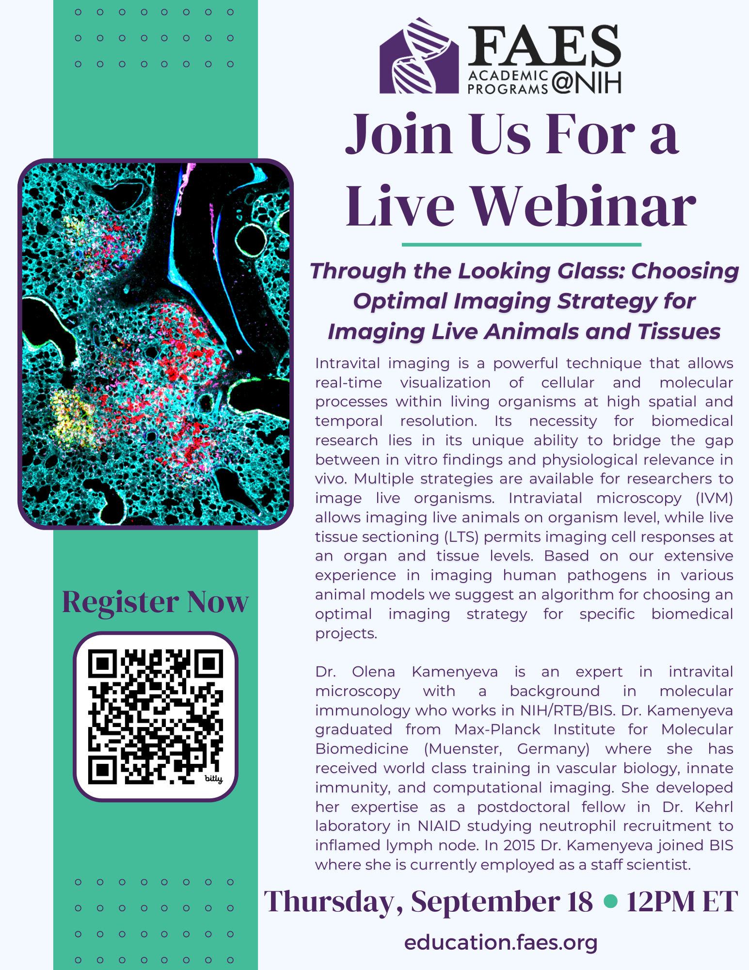
FAES Educational Webinar Series: Through the Looking Glass: Choosing Optimal Imaging Strategy for Imaging Live Animals and Tissues
Intravital imaging is a powerful technique that allows real-time visualization of cellular and molecular processes within living organisms at high spatial and temporal resolution. Its necessity for biomedical research lies in its unique ability to bridge the gap between in vitro findings and physiological relevance in vivo. Multiple strategies are available for researchers to image live organisms. Intraviatal microscopy (IVM) allows imaging live animals on organism level, while live tissue sectioning (LTS) permits imaging cell responses at an organ and tissue levels. Based on our extensive experience in imaging human pathogens in various animal models we suggest an algorithm for choosing an optimal imaging strategy for specific biomedical projects. Those who are interested in learning more about this fascinating topic are encouraged to enroll in the FAES workshop Intravital Microscopy for Biomedical Research (BIOC 036).
Dr. Olena Kamenyeva is an expert in intravital microscopy with a background in molecular immunology who works in NIH/RTB/BIS. Dr. Kamenyeva graduated from Max-Planck Institute for Molecular Biomedicine (Muenster, Germany) where she has received world class training in vascular biology, innate immunity, and computational imaging. She developed her expertise as a postdoctoral fellow in Dr. Kehrl laboratory in NIAID studying neutrophil recruitment to inflamed lymph node. In 2015 Dr. Kamenyeva joined BIS where she is currently employed as a staff scientist. Dr. Kamenyeva is also teaching Intravital Microscopy for Biomedical Research (BIOC 036) in the FAES Learning Labs this October.
Return to News / Events